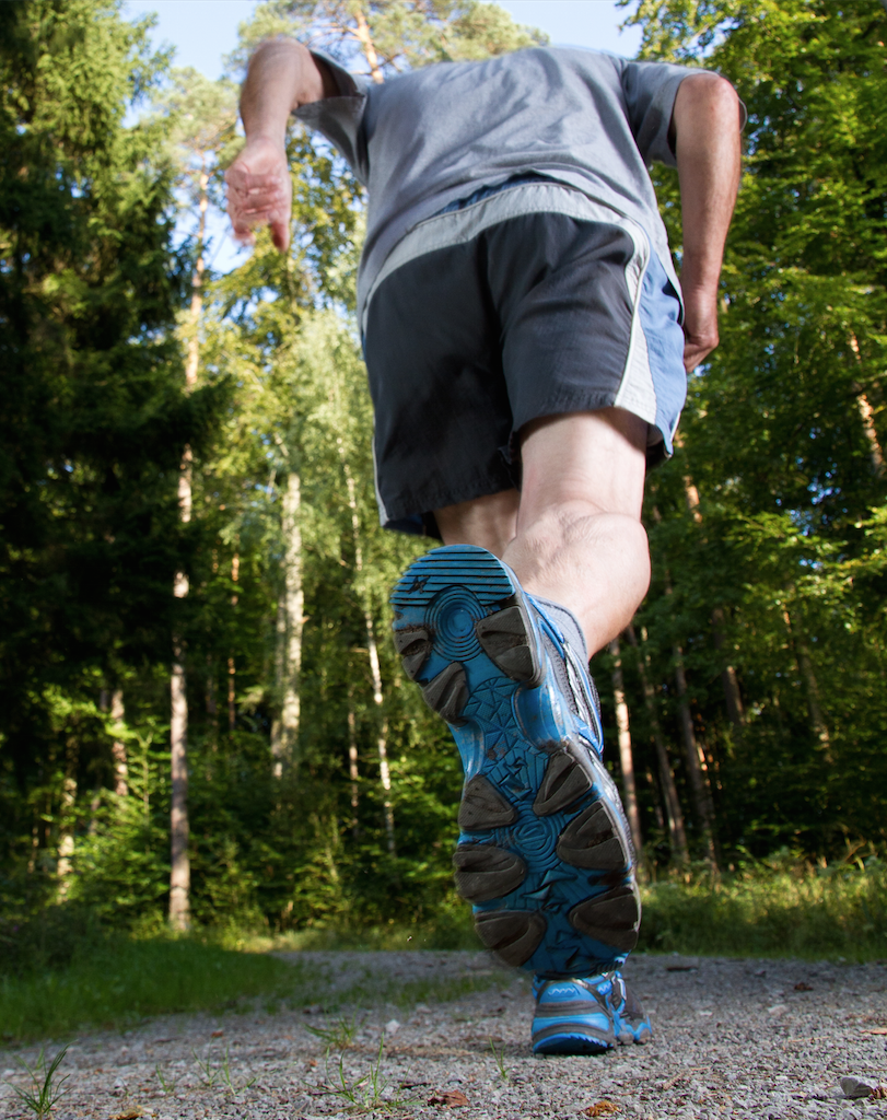
Getting runners back on the run after heel pain can be challenging at times. Accordingly, this author offers key diagnostic insights, reviews a variety of possible etiologies ranging from biomechanical causes to nerve-related heel pain, and shares his clinical experience on viable treatment options.
Heel pain is one of the most common foot ailments that runners experience. Not only are runners affected physically but many of them also suffer psychologically due to the loss of an important routine in their lives. In order to treat the pain correctly, we must first determine the etiology and mechanism of the disorder.
Accordingly, let us take a closer look at the most common causes of heel pain along with treatment options. One thing to remember in treating anyone with heel pain is that everyone responds differently to treatment protocols.
Even today, some physicians tell patients they have heel spurs. In actuality, the presence or absence of a plantar calcaneal spur often has little to do with the cause of the pain except in rare cases. Historically, we also thought that inflammation of the plantar fascia is what caused the pain. We now know the pain is more due to chronic thickening of the plantar fascia.
Clinicians can use X-rays to rule out other causes of heel pain. Musculoskeletal ultrasound has been an excellent diagnostic tool as it allows one to measure the thickness of the plantar fascia against the norm. Runners usually complain of pain being the worst when they first get up in the morning or after other periods of rest. With activities, the pain subsides although prolonged standing/ambulation on hard surfaces may cause increased pain. The pain is usually the worst at the medial band of the plantar fascia at the attachment into the medial plantar calcaneal tubercle.
Tightness of the gastrocnemius soleus/Achilles tendon complex in theory is the main cause of painful heels not only in runners but also in the general population. While this tightness is present the majority of the time, there can be other muscle issues with or without the presence of equinus. Often, tightness of the hamstrings may cause early heel off in runners. One must address these issues in order to relieve pain and minimize recurrence. Evaluation of the entire kinetic chain helps in designing a stretching and strengthening program for the runner.
A Closer Look At Treatment Options For Plantar Fasciitis
Another area to evaluate is the biomechanics of the runner. Some over-the-counter/prefabricated orthotics can provide a temporary or permanent solution to control the amount and speed of pronation. Prefabricated orthoses are a less expensive option if insurance does not cover custom orthoses. In other cases, a custom orthotic is necessary with various modifications. An extended forefoot varus post helps to control pronation once heel off occurs and forefoot loading takes place.
 If a runner has functional hallux limitus, a reverse Morton’s extension with a first ray cutout and posting for metatarsals two through five will allow for more first metatarsal plantarflexion, thus enabling more dorsiflexion of the hallux. One can also use a Cluffy Wedge (Cluffy) under the hallux to preload in dorsiflexion, again allowing for increased motion. For a more rigid first metatarsophalangeal joint (MPJ), I will use a semi-rigid to rigid Morton’s extension, attaching it either to the orthotic or most often on a separate graphite plate that I place under the orthotic.
In patients with a hypermobile rearfoot, deep heel cups, high phalanges and wider orthotics will increase control. Heel lifts can temporarily reduce the strain on the Achilles complex but one should reduce or remove the heel lift as the patient progresses with the stretching program. One can also modify the cast for the orthotics to increase their effectiveness. The patient can plantarflex the first ray without a fill for metatarsals two through five on the cast. Clinicians can also order the medial longitudinal arch fill as minimal, normal or maximal, or with no fill.
For many years, one of the first-line treatments has been steroid injection. While this can be helpful to reduce the pain and thickness of the plantar fascia, there are also risks with these types of injections. Fat pad atrophy, spontaneous rupture of the plantar fascia, neuropraxia of plantar nerves, the steroid flare/rebound effect and failure to relieve the pain are just some of the negative consequences. Recent injection alternatives include platelet-rich plasma or liquid amniotic tissue. Ultrasound-guided injections are becoming more popular to direct the treatment more accurately in the area of maximum pathology. Due to stress on the plantar fascia in running, one should minimize the use of injections and use them after the failure of non-invasive therapies.
Most of the time, I will refer patients to physical therapy. Good physical therapists are worth their weight in gold. Not only will they help evaluate the musculoskeletal system along with the kinetic chain, they can prescribe the proper stretching and strengthening exercises. In addition, modalities such as ultrasound, electrical stimulation, iontophoresis, ice massage and cold laser have been successful adjunct treatments in my experience. With increased thickness of the plantar fascia in most cases, deep tissue massage with and without instruments can reduce the thickness and pain. Patients have shown some progress with low-energy shockwave treatments. These usually occur in a doctor’s office with no anesthesia although one can perform them in a home or outpatient clinic. Multiple treatments are usually necessary.
 We often overlook the evaluation and modification of external factors but they can be vitally important. The midsole of most running shoes will only last 300 to 500 miles depending on the softness of the midsole. The rest of the shoe may look fine but the function of the midsole is greatly decreased. This can lead to overuse pain or a change in biomechanics. Purchasing new shoes may help to eliminate some or even all of the pain.
One should question all runners about their training habits. This includes terrain, speed, surface type and body weight. Would the athletes be able to run with less pain if they switched from hills to flatter terrain? How would soft surfaces like grass compare to asphalt or concrete? Another factor would include running on the side of a hill versus on a flat surface. Weight loss will help reduce the stress on the body during gait. Considering that running places three to five times the body’s weight on the lower extremities, the heavier a runner, the more likely he or she will develop lower extremity overuse syndromes. Plantar fascia night braces keep the muscles from shortening in the foot and calf areas. This is especially helpful in relieving post-static dyskinesia upon arising in the morning.
When Should You Consider Surgery For Plantar Fasciitis In Runners?
Surgery should only be a last resort, especially in runners. Prior to invasive surgery, I have used high-energy shockwave therapy. Patients are usually under sedation in an outpatient setting. The high-energy machine usually only requires one treatment but does impact the area with more force than low energy ESWT. Also, due to the noise of the machine, ear protection is mandatory.
Traditional open surgery involves release of the plantar fascial insertion from medial to lateral, most often through a medial or plantar incision. Some surgeons choose to remove the underlying bone spur if it is present. This surgery traditionally required a long recovery period, which is not a good choice for runners. More recently, surgery has evolved around minimal incision type of procedures. The in-step procedure entails a small incision, usually between the heel and the medial longitudinal arch. In most patients, this is in a non-weightbearing area of the foot. One would only release the medial band or remove a section of the medial band to allow for further lengthening of the fascia.
One of the biggest advancements in foot surgery has been the advent of endoscopic procedures. Endoscopic plantar fasciotomy has reduced the recovery period for many patients. The patient can bear weight immediately since the incisions are small and located medially and laterally on the heel. Similar to performing the in-step procedure, surgeons should take care to release the medial band only. A non-weightbearing lateral X-ray is necessary for preoperative planning. There is also a learning curve for the procedure.
Key Insights On Calcaneal Stress Fractures
When runners describe their pain as getting worse the more they are on their feet, one has to be concerned about the possibility of a calcaneal stress fracture. One can elicit pain with side-to-side compression of the calcaneus, palpation of the medial and lateral plantar heel, and palpation of the medial and lateral walls of the calcaneus away from the medial calcaneal and sural nerve branches. X-rays may or may not show any changes. In some cases, a white sclerotic line will be visible on the calcaneus. Further imaging studies such as a bone scan or a magnetic resonance image (MRI) may be needed.
Often, the cause of the stress fracture is a sudden increase in training/mileage or speed. Other known causes are worn out shoes (too many miles), increased body weight, a change in surface or a change in terrain. Clinicians should also evaluate runners with possible stress fractures for low vitamin D levels.
Treatment revolves around controlling the pain. After diagnosing a stress fracture, apply a below-knee cast or at least a controlled ankle motion (CAM) walker. Non-weightbearing will speed up the healing time by eliminating the plantar pressure altogether. When athletes wear a cast and/or crutches for four to six weeks, they will often develop muscle atrophy so early physical therapy is necessary to prevent loss of strength. Sometimes patients will start off with plantar fasciitis, continue to run and then develop a stress fracture. When the fracture pain ends, they may revert back to having plantar fasciitis pain. After the fracture, they may need follow-up treatment for the plantar fasciitis as I have outlined previously. One can employ orthotics to manage faulty biomechanics and new running shoes may also be necessary. Evaluate the athlete’s training to look for possible mitigating factors. The return to running should be gradual with lower mileage along with softer and flatter courses.
When Inflammatory Arthritis Causes Heel Pain
Rheumatoid arthritis and seronegative spondyloarthropathies can be causes of pain in the heels. Most of the time, patients will also complain of back pain as the disease process causes pain in the heels and back. The pain is usually diffuse but tends to follow the back of the calcaneus. Post-static dyskinesia may be present as well.
The pain has a local and a systemic manifestation so one should treat both. Local treatment includes orthotics (soft or semi-rigid), a change in shoes, control of faulty mechanics and a change in training. For systemic treatments, I refer the patient to a rheumatologist for medical management and a physical therapist for physical therapy.
Treating Nerve-Related Heel Pain
For some patients, the source of heel pain emanates from the nervous system. The most common of these conditions is tarsal tunnel syndrome, which is an entrapment of the posterior tibial nerve in the medial neurovascular bundle around the tibial malleolus. The etiology is often mechanical due to increased pronatory forces that cause a stretch on the nerve. Other known causes have been soft tissue masses such as ganglionic or fibrous cysts as well as lipomas. Varicose veins of the greater saphenous vein can also wrap around or cause pressure on the nerve. The runner will describe the pain as sharp, shooting, burning and/or radiating either superior or inferior.
X-rays may show an increased soft tissue density. There is usually a positive Tinel’s sign with radiating pain when tapping or pressing on the nerve in the tarsal tunnel area. While an electromyogram/nerve conduction velocity (EMG/NCV) test has been the traditional diagnostic test, it is not always accurate and can be very painful for the patient. A MRI may detect soft tissue masses or other abnormalities in the tarsal tunnel area. Injection of a local anesthetic into the tarsal tunnel area can also be diagnostic if it totally relieves the pain.
 Non-surgical treatment includes orthotics to control abnormal pronation. If a soft tissue mass is causing pain, an ultrasound-guided injection will help with the placement of a steroid directly into the soft tissue mass. Physical therapy has had limited success but iontophoresis and electrical stimulation can reduce the pain. If conservative treatment fails and pain persists, surgery may be needed. This would consist of decompression of the tarsal tunnel and removal of any soft tissue cysts or varicose veins. Dissection should continue down into the foot, freeing the medial and lateral plantar nerves along with their branches down into the foot behind the abductor hallucis muscle. Patients will need physical therapy postoperatively to prevent deep scar tissue, which could cause recurrence of the symptoms.
Similar to tarsal tunnel syndrome, there are two other areas where nerves can be entrapped. One is the medial calcaneal nerve or Baxter’s nerve. This branch penetrates through the abductor hallucis muscle where it gets entrapped. The pain usually is on the medial heel with some superior radiating pain. That nerve does seem to respond to alcohol sclerosing injections and physical therapy such as iontophoresis. The medial and lateral plantar nerves can also be entrapped superior or inferior to the abductor hallucis muscle. This is in a different area than the tarsal tunnel. Release of the nerve would involve releasing the fascia of the abductor hallucis muscle superiorly and inferiorly.
Another cause of heel pain may be radiculopathy from the low back. Most of the heel is innervated by the S2 disc with surrounding areas innervated by the L4, L5 and S1 discs. Bulging discs, arthritis, spondylolisthesis and other impingement or narrowing problems can cause radiating pain to the heel. X-rays of the back may or may not show pathology. In these cases, a MRI or computed tomography (CT) scan is necessary. Give the patient a referral to physical medicine and rehabilitation and/or neurosurgery for evaluation and management. Usually, this consists of physical therapy and/or epidural injections. Since surgery may be quite debilitating to the runner, do it only after exhausting conservative treatment.
Pertinent Pearls On Managing Posterior Heel Pain
Pain in the posterior aspect of the heel can be difficult to treat in many athletes. There are two types of posterior deformities in this area. The first is the traditional Haglund’s deformity. This is usually a prominence of the superior, posterior lateral aspect of the calcaneus. The etiology can be structural due to varus rotation of the heel or a high calcaneal inclination angle. Other causes include muscle tightness, trauma, bone cysts or tumors, and excessive pronation with irritation to the Achilles tendon. The counter of the shoe is often too rigid so there is pressure from the shoe on the calcaneus. This can cause bursitis as well as thickening of the Achilles tendon.
Non-surgical treatment consists of removing the irritating force from the calcaneus and/or controlling excessive pronation. Shoe modification involves softening of a rigid heel counter or switching to shoes that have a softer heel counter. One can place padding in the shoes as well as heel cups to reduce shearing forces. There are elastic ankle braces with silicone or gel padding along the posterior heel area that reduce pain. If there is excessive pronation, one needs to control this with a foot orthotic. Stretching and strengthening exercises along with iontophoresis can reduce the pain as well.
Non-steroidal anti-inflammatory drugs or oral prednisone may reduce pain and inflammation. Use injections with great caution in this anatomic area. There have been cases of partial or complete rupture of the Achilles tendon, especially with steroid injections. Instead, I recommend platelet rich plasma injections or even amniotic tissue in injectable form under ultrasound guidance.
 Surgery could involve just removing the superior, posterior, lateral aspect of the calcaneus. This usually does not involve much dissection or release of the Achilles tendon. Some surgeons may perform this as an endoscopic procedure. However, if the etiology is due to varus deformity or an increased calcaneal inclination angle, a calcaneal osteotomy may be necessary. Usually, if you only remove the bump, the patient is non-weightbearing for two weeks with four subsequent weeks in a CAM walker. Aggressive physical therapy starts around three to four weeks. However, if one performs an osteotomy, the patient is usually non-weightbearing for six to eight weeks with physical therapy to follow.
There is also posterior heel pain that can occur at the insertion of the Achilles tendon. This is usually due not only to shoe irritation but tightness of the Achilles tendon and/or hamstring. It is located more plantarly than a Haglund’s deformity when looking at the posterior heel. This area is very different than the Haglund’s deformity. There is an enthesopathy of the insertion of the Achilles tendon. The calcification that forms is on the anterior aspect of the Achilles tendon and is often embedded in the tendon fibers.
Conservative treatment is much the same with the exception of steroid injections for both causes of posterior heel pain and one should exhaust conservative care first. With the enthesopathy, detach the Achilles tendon or have it flapped excessively. Remove the calcified area, repair the tendon and reattach it using bone anchors. The patient is non-weightbearing for four to six weeks. Subsequently, the patient wears a CAM walker and has physical therapy for another four to six weeks.
In Summary
When it comes to runners, it is vital to fully understand the etiology and treatment of heel pain in order to have better outcomes and adherence from the patient. It is also important that if the athlete will be unable to run that you recommend an alternative exercise program to maintain fitness. This could include running in the deep end of a pool with a flotation belt, use of a stationary bike or upper body weight training. Always exhaust conservative treatment options before contemplating surgery.
If you believe you may be experiencing the painful symptoms of Runner’s Heel Pain, contact Achilles Podiatry for further evaluation and treatment.
Source: Podiatry Today

